
Article
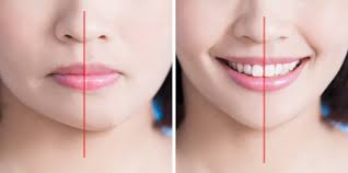
Fixed Orthodontic Appliances and Early Caries: Risk Factors and Preventive Strategies
Fixed orthodontic appliances have transformed smiles, but they also create ideal conditions for Initial Caries Lesions (ICLs), especially white spot lesions around brackets. Research shows their prevalence can range from 27% to 97%, largely driven by hygiene and compliance1.
For dentists, prevention must begin the day appliances go on. Quick risk screening, strong hygiene support, and early enamel-protective interventions are key to stopping minor demineralization from turning into lasting damage.
Risk Factors for Early Caries
1. Plaque Retention and Biofilm Accumulation
Fixed orthodontic appliances increase plaque buildup as brackets, bands, and wires hinder natural self‑cleaning and complicate brushing. These retention sites promote biofilm accumulation, lowering pH, and tipping the balance toward demineralization. If persistent, this process leads to initial carious lesions and, eventually, cavitated defects 1.
2. Altered Salivary Dynamics
Fixed appliances interfere with natural salivary flow and buffering capacity. Reduced clearance of fermentable carbohydrates prolongs acid exposure, accelerating lesion initiation.
3. Dietary Habits
Orthodontic patients are often advised to eat softer foods, but many turn to convenient options like pastries, cookies, ice creams, and carbonated drinks. These sugar‑rich choices, combined with plaque retention around brackets and bands, lower pH and promote enamel decalcification, which can progress to carious lesions if uncontrolled2.
4. Poor Oral Hygiene Practices
Maintaining consistent oral hygiene can be challenging for individuals with fixed orthodontic appliances. The presence of brackets, wires, and bands makes plaque removal more difficult, and irregular brushing may contribute to plaque accumulation. When oral hygiene is not adequately maintained, the risk of demineralization and the development of early caries lesions increases3.
5. Appliance‑Related Challenges
Fixed orthodontic appliances can increase caries risk due to plaque accumulation around brackets, wires, and bands. These attachments disrupt natural self‑cleansing and make brushing more difficult4.
5. Diagnostic Blind Spots
Traditional visual and radiographic methods often miss early proximal lesions. Without adjunctive imaging, lesions may progress unnoticed until cavitation occurs.
Preventive Strategies for Practicing Dentists
1. Reinforced Oral Hygiene Protocols
- Dentists should emphasize meticulous brushing with fluoride toothpaste, supplemented by interdental brushes and floss threaders3.
- Chairside demonstrations and motivational interviewing can improve compliance.
2. Topical Fluoride and Remineralization Agents
- Regular application of fluoride varnishes, gels, or rinses strengthens enamel against acid attack.
- Casein phosphopeptide‑amorphous calcium phosphate (CPP‑ACP) and nano‑hydroxyapatite formulations further enhance remineralization5.
3. Dietary Counseling
- Encouraging patients to limit sugary snacks and acidic beverages is essential.
- Advising water rinses after meals and promoting sugar‑free chewing gum can help neutralize acids6.
4. Advanced Diagnostic Tools
- Near‑infrared imaging (NIRI) and intraoral scanners allow early detection of proximal lesions invisible to conventional methods7.
- Incorporating these technologies into routine orthodontic monitoring can prevent progression.
5. Sealants and Protective Coatings
- Applying resin sealants or protective coatings around brackets reduces enamel exposure to cariogenic biofilm. This strategy is particularly effective in high‑risk patients with poor hygiene.
6. Patient Education and Motivation
- Visual aids showing white spot lesions can be powerful motivators.
- Regular reinforcement of hygiene instructions and positive feedback during appointments encourage long‑term compliance.
Final Takeaway
Fixed orthodontic appliances reshape more than smiles—they reshape the oral environment. Without proactive prevention, enamel health is at risk. The winning formula for clinicians is clear: combine fluoride, smart diet guidance, advanced diagnostics, and patient education to keep braces esthetic and functional without sacrificing enamel integrity.
Reference
- Salerno C, Cagetti MG, Cirio S, Esteves-Oliveira M, Wierichs RJ, Kloukos D, Campus G. Distribution of initial caries lesions in relation to fixed orthodontic therapy: a systematic review and meta-analysis. Eur J Orthod. 2024;46(2):cjae008. doi:10.1093/ejo/cjae008.
- Ali M, Nahidh M. Diet and orthodontics: a review. J Baghdad Coll Dent. 2021;33(3):30-38. doi:10.26477/jbcd.v33i3.2951.
- Almutairi RM, Alturaif DJ, Alanzi LM. Importance of oral hygiene in orthodontic treatment. Saudi J Oral Dent Res. 2023;8(3):100-9.
- ElNaghy R, El Sayed M, Alnaghy RA, Hasanin M. Does orthodontic treatment using fixed appliances cause initial caries lesions?. Evidence-Based Dentistry. 2024 Sep;25(3):134-5.
- Rane GA, Sandhyarani B, Patil AT, Keshaw K, Raikar A, Davalbhakta R. Comparative evaluation of the depth of remineralization with casein phosphopeptide-amorphous calcium phosphate, sodium fluoride with CXP, nano-hydroxyapatite, and sodium fluoride on artificially demineralized lesions using stereomicroscope: an in vitro study. Journal of Indian Association of Public Health Dentistry. 2023 Apr 1;21(2):118-23.
- Hattab FN. Chewing gum for oral and dental health: a review. Journal of Oral Health and Community Dentistry. 2023 Jan;17(1):13.
- Michou S, Vannahme C, Bakhshandeh A, Ekstrand KR, Benetti AR. Intraoral scanner featuring transillumination for proximal caries detection. An in vitro validation study on permanent posterior teeth. Journal of Dentistry. 2022 Jan 1;116:103841.
Related Contents
Video
Management of Traumatic Tooth Avulsion: When & How?
Traumatic tooth avulsion is a true dental emergency requiring immediate and structured management to...
Article
Prosthetic Design Factors That Prevent Peri-Implant Bone Loss
You placed the implant perfectly. Osseointegration was flawless. But three years later, there is 3mm...
Article
Treatment Algorithms for Peri-Implant Mucositis and Peri-Implantitis
You can prevent most peri-implant disease. But once it develops, treatment becomes less predictable....
Article
Implant Surface Decontamination: What Actually Works
Treating peri-implantitis starts with one critical step, which is decontaminating the implant surfac...
Article
Growth & Facial Asymmetry: When to Worry
Only a few dentists can notice it and realize the asymmetry can signal an underlying skeletal imbala...
Article
Orthodontic Red Flags Every Dentist Should Recognize: Functional Habits and Airway Cues
Some malocclusions cases stall for reasons you can’t see on a scan, as not...
Article
Tooth Eruption & Space Management: What Orthodontists Should Watch For
Tooth eruption may not alwaxfys fit the predictable biological timeline. A delayed or ecto...
Article
Communication & Referral Timing: Getting It Right
You have identified a problem, maybe a severe Class III malocclusion in a 9-year-old, or it is an im...