Article
Managing Common Oral Mucosal Lesions: A Practical Guide for Dentists
Most dental work is related to restorations, extractions, and routine care, but a sizable portion comes with oral mucosal complaints.
For dentists, it is critical to spot and manage common mucosal conditions in daily practice.
They should be able to recognise what looks benign, treat what is straightforward, and make an urgent referral when required.
Here is the practical guide to help you with every step.
Why mucosal care matters in primary dental practice?
Oral mucosal disease management is somewhat in the middle of medicine and dentistry.
Lesions in the mouth can be1:
Local in the form of trauma or denture-related injuries
Infectious, like oral candidiasis or herpetic lesions
Immune-mediated, such as oral lichen planus
Systemic due to nutritional deficiencies or Crohn’s
Potentially malignant, like leukoplakia or erythroplakia
Oral diagnosis remains fundamental for patients for a better prognosis. However, due to limited access, biopsies and further treatment is usually conducted by medical professionals.
A practical chairside triage (quick checklist)
- Duration - If ulceration extends beyond 3 weeks, you should consider referring the patient for biopsy2.
- Appearance - Persistent red or mixed red-white patches are at higher risk than homogeneous white plaques.
- Location & behavior - Lesions on the lateral tongue, floor of the mouth, and lesions that are indurated, fixed, or bleed easily should be addressed on an urgent basis1.
- Predisposing factors - Consider predisposing factors like prolonged denture wear, xerostomia, diabetes, immunosuppression, tobacco/alcohol use3. Try to correct or address these when possible.
Common conditions: What to do?
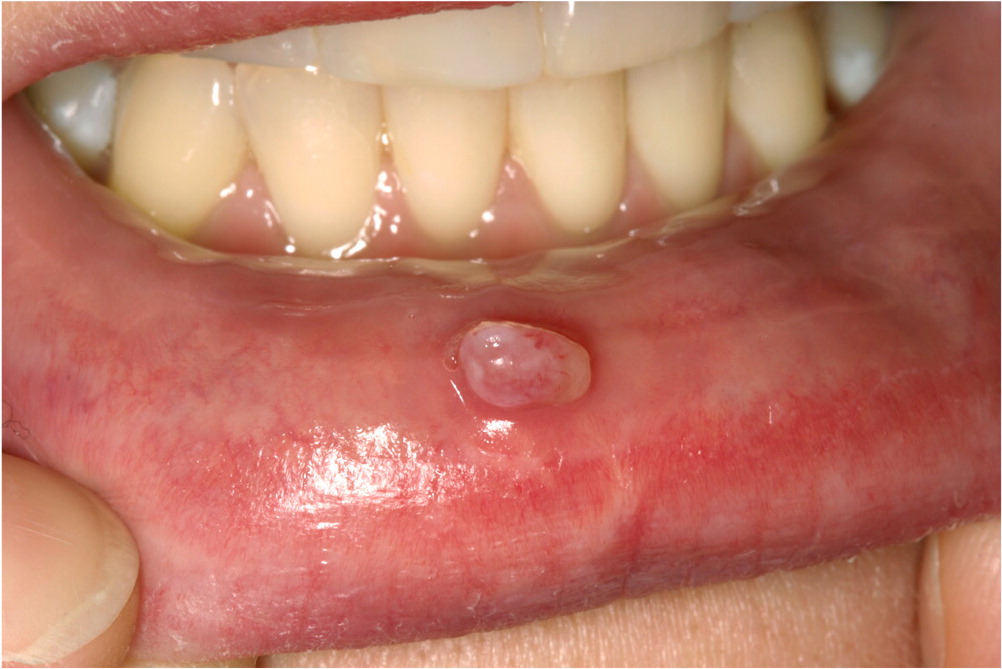
1. Recurrent aphthous stomatitis (RAS)
It is seen as small, round/oval, painful ulcers on non-keratinised mucosa.
Identify local trauma and obvious triggers for the condition. Use topical corticosteroids for symptomatic relief. You can also suggest the use of protective pastes and analgesic gels.
If ulcers are unusually large or are accompanied by some systemic symptoms, refer to an appropriate medical professional.
2. Oral candidiasis (denture stomatitis, thrush)
It is seen as wipeable white plaques, erythematous mucosa, or angular cheilitis.
Oral candidiasis treatment for dentists includes using topical antifungals and ensuring denture hygiene. Redo instructions and ask the patient to clean and soak the denture overnight.
Check for predisposing causes like poorly-controlled diabetes, xerostomia, or use of recent antibiotics3.
Topical therapies remain the first course of action, go on to antifungal medications for the extensive stage.
3. Oral Lichen Planus (OLP)
It is seen as reticular white striae, which are often asymptomatic. There are also erosive/ or atrophic forms, which can be painful.
For asymptomatic reticular lesions, only observe and document. For symptomatic lesions, start with topical corticosteroids and manage secondary candidal infection if present.
Arrange regular reviews in case of erosive oral lichen planus, as it carries a small malignant-transformation risk. Therefore, long-term surveillance is recommended4.
Refer to a specialist if there is no improvement for further investigation.
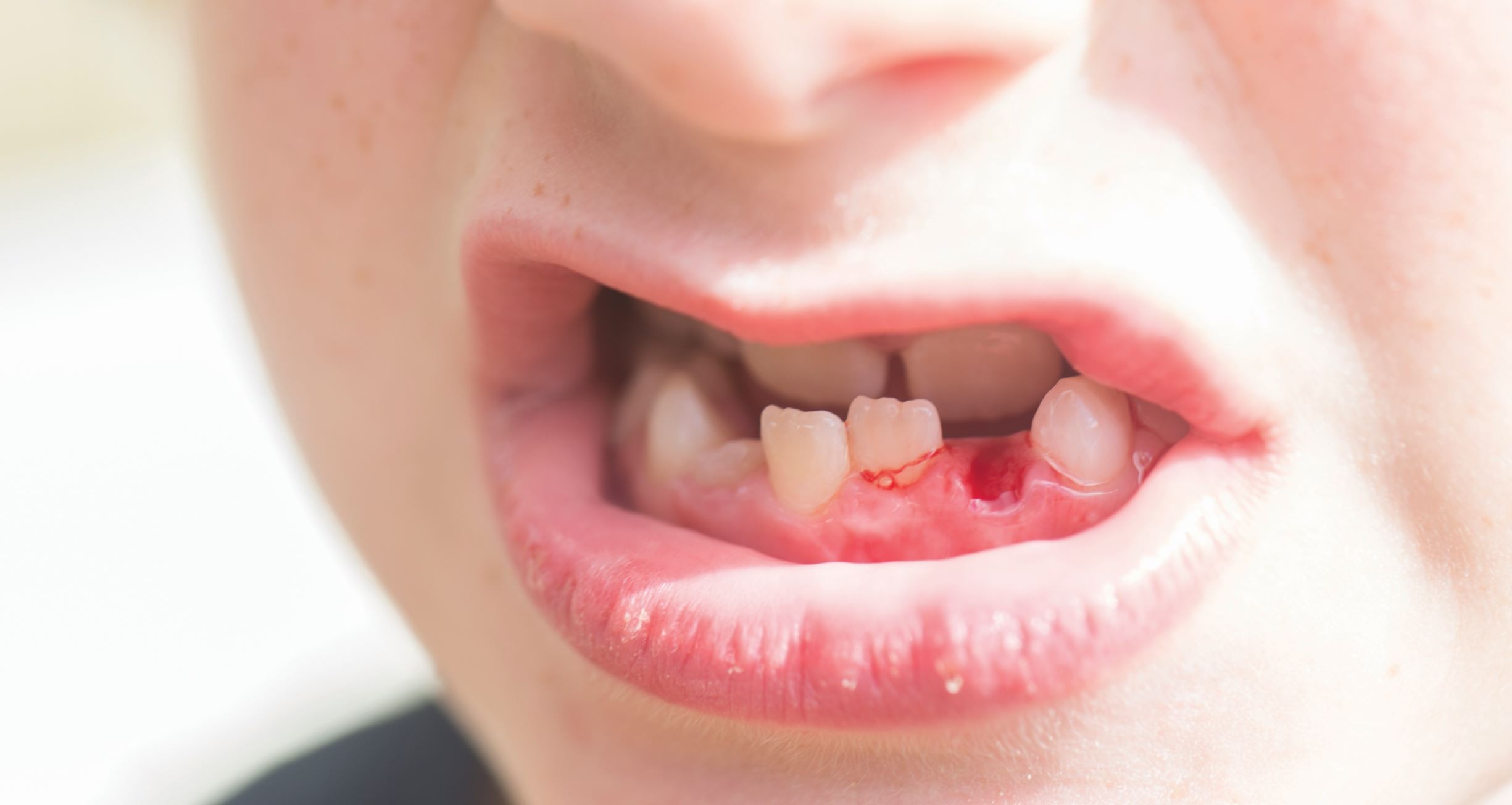
4. Traumatic and frictional lesions
It can be seen as well-localised white patches or ulcers adjacent to sharp teeth, restorations, or ill-fitting prostheses.
It is important to identify and remove the source. You can adjust or smooth sharp edges, or reline/repair a faulty prosthesis.
Provide topical analgesia if needed. Healing is expected within ~7–10 days; if not healed after removal of the cause, refer to a specialist.
5. Leukoplakia, erythroplakia, suspicious patches
It is seen in the form of persistent white or red patches that cannot be attributed to a known cause, and are leukoplakia and erythroplakia, respectively5.
Try to exclude reversible causes like tobacco use or traumatic keratosis.
If a patch persists beyond 2–3 weeks and is non-homogeneous or indurated, refer to a specialist for biopsy.
Erythroplakia in particular has a high malignant potential.
Documentation
It is important to record lesion size, site, morphology and duration and document it. Click photographs of lesions with good lighting.
They can help when you consult a specialist and also compare the lesions in upcoming follow-up.
Red flags that need urgent action
- Unexplained oral ulceration > 3 weeks.
- Persistent red or mixed red-white patches (erythroplakia/erythroleukoplakia).
- A firm/indurated lump, rapidly enlarging lesion, or persistent bleeding.
Final Takeaway
For mucosal lesions in the mouth, consider the duration, appearance, and site, along with risk factors.
Consider topical therapy and rectify any local causes. Always document the lesion at every visit and refer the patient in case of any red flags. Do not ignore any lesion, and remember that earlier biopsy and diagnosis can save lives.
References
- Atkin PA, Cowie R. Oral mucosal disease: dilemmas and challenges in general dental practice. Br Dent J. 2024 Feb 23;236(4):269-273.
- NICE Guideline NG12 (2025). Suspected cancer: recognition and referral. https://www.nice.org.uk/guidance/ng12
- Taylor M, Brizuela M, Raja A. Oral Candidiasis. In: StatPearls [Internet]. Treasure Island (FL): StatPearls Publishing; 2023 Jul 4.
- Manchanda Y, Rathi SK, Joshi A, Das S. Oral Lichen Planus: An Updated Review of Etiopathogenesis, Clinical Presentation, and Management. Indian Dermatol Online J. 2023 Dec 22;15(1):8-23.
- Öhman J, Zlotogorski-Hurvitz A, Dobriyan A, Reiter S, Vered M, Willberg J, Lajolo C, Siponen M. Oral erythroplakia and oral erythroplakia-like oral squamous cell carcinoma – what’s the difference? BMC Oral Health. 2023 Nov 13;23:859.
Related Contents
Video
Management of Traumatic Tooth Avulsion: When & How?
Traumatic tooth avulsion is a true dental emergency requiring immediate and structured management to...
Article
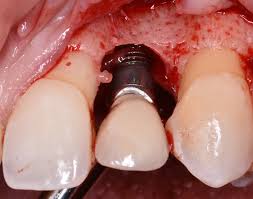
Prosthetic Design Factors That Prevent Peri-Implant Bone Loss
You placed the implant perfectly. Osseointegration was flawless. But three years later, there is 3mm...
Article
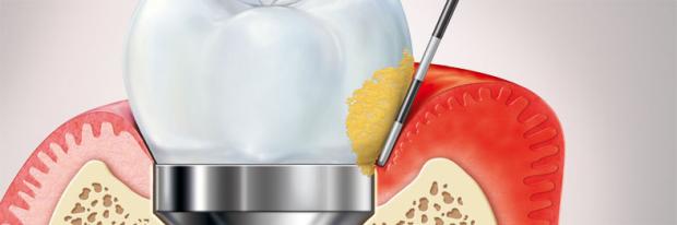
Treatment Algorithms for Peri-Implant Mucositis and Peri-Implantitis
You can prevent most peri-implant disease. But once it develops, treatment becomes less predictable....
Article
Implant Surface Decontamination: What Actually Works
Treating peri-implantitis starts with one critical step, which is decontaminating the implant surfac...
Article
Growth & Facial Asymmetry: When to Worry
Only a few dentists can notice it and realize the asymmetry can signal an underlying skeletal imbala...
Article
Orthodontic Red Flags Every Dentist Should Recognize: Functional Habits and Airway Cues
Some malocclusions cases stall for reasons you can’t see on a scan, as not...
Article
Tooth Eruption & Space Management: What Orthodontists Should Watch For
Tooth eruption may not alwaxfys fit the predictable biological timeline. A delayed or ecto...
Article
Communication & Referral Timing: Getting It Right
You have identified a problem, maybe a severe Class III malocclusion in a 9-year-old, or it is an im...