
Article
Dental Trauma Guide 2025: Managing Avulsion, Luxation & Fracture
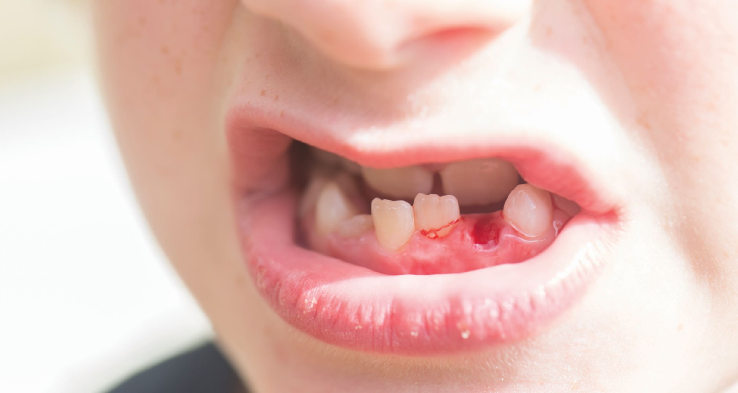
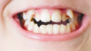
Dental trauma is a common incident and can happen during sports, accidents, or even routine play.
The first few minutes after an injury determine whether a tooth can be saved or lost.
This guide outlines evidence-based chairside emergency protocols for avulsion, luxation, and fracture aligned with the latest IADT 2020 Guidelines, AAPD, and AAE trauma recommendations1,2.
Step 1: Assessment
Before you reach for instruments, stabilize the situation.
Key Assessment Checklist:
- Find out the time since injury, since it is critical for prognosis.
- Tooth condition and look if the tooth is avulsed, luxated, or fractured.
- Soft tissue & socket and check for embedded fragments or alveolar fractures.
- Consider medical factors and find out if there was any loss of consciousness, and if the tetanus shot is up to date.
- Take two angled periapicals or CBCT if root fracture suspected.
Clinical Tip:
Always assess adjacent and opposing teeth. Secondary trauma is quite common but is often missed in the first exam3.
Step 2: Avulsion Management
Avulsion is a true dental emergency. Prognosis depends entirely on how long the tooth stays dry1.
Golden 15-Minute Window
- Replant immediately if <15 minutes passed, at the site if possible.
- If contaminated, you can rinse gently with saline or milk. Never scrub the root surface.
- Storage media (in order of preference):
- Hank’s Balanced Salt Solution (HBSS)
- Cold milk
- Saline
- Patient’s saliva. Store in buccal vestibule if conscious and cooperative
Chairside Replantation Protocol
- Handle the crown only and avoid touching the root.
- Irrigate the socket with saline and remove the visible clot gently.
- Replant using light finger pressure.
- Confirm position radiographically.
- Apply flexible splint for 2 weeks. You can extend to 4 weeks if the dry time >60 min or an alveolar fracture is seen.
- Prescribe systemic antibiotics like amoxicillin or doxycycline.
- Update tetanus status if needed.
Endodontic Management
- Closed apex: Begin RCT 7–10 days post-replantation; before splint removal.
- Open apex: Monitor for revascularization before intervening.
Important note: Never replant avulsed primary teeth, as it increases the risk of damaging developing permanent buds.
Step 3: Luxation Injuries
Luxation includes a range of partial displacements. Quick repositioning and stabilization are key to saving the tooth4.
|
Type |
What You See |
Recommendation |
|
Concussion / Subluxation |
Tenderness, no displacement |
Soft diet, occlusal adjustment, monitor vitality |
|
Extrusive / Lateral Luxation |
Elongated or shifted tooth |
Gently reposition under LA, flexible splint for 2–4 weeks |
|
Intrusive Luxation |
Tooth pushed into socket |
Open apex: allow spontaneous re-eruption (up to 3 weeks) Closed apex: orthodontic or surgical repositioning + RCT after 2 wks |
Chairside Tip:
Use gentle digital pressure for repositioning without forcing it against alveolar resistance. Take post-reposition radiographs to check socket integrity.
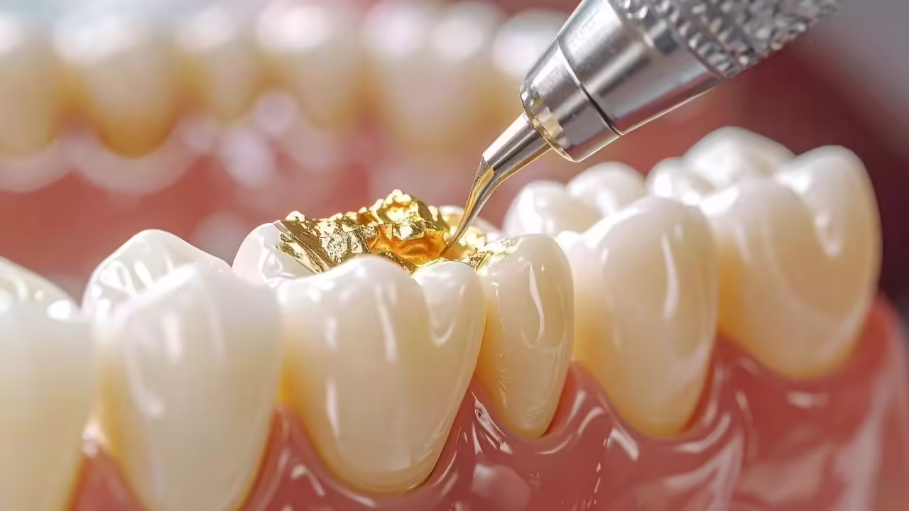
Step 4: Managing Tooth Fractures
Fractures of teeth are common but manageable when approached methodically.
Enamel–Dentin Fracture
- Cover exposed dentin with calcium hydroxide or resin-modified glass ionomer.
- If a fragment is available, store it in saline and reattach.
Crown–Root Fracture
- Stabilize temporarily with fiber splint.
- Assess restorable margin and later on manage with crown lengthening or orthodontic extrusion.
Root Fracture
- Confirm with multiple angulated X-rays or CBCT.
- Reposition the coronal fragment, splint 4–6 weeks.
- Initiate RCT only if pulp necrosis develops.
Clinical Insight:
Always record mobility, color changes, and pulp response. These form your baseline for follow-up and medicolegal protection.
Step 5: Splinting & Follow-Up
Here is the protocol for splinting in different situations, along with the recommended follow-up schedule.
|
Condition |
Recommended Splint Duration |
Material |
|
Avulsion |
2–4 weeks |
0.4 mm wire + composite (flexible) |
|
Luxation |
2–4 weeks |
Flexible wire or fiber |
|
Root fracture |
4–6 weeks |
Fiber-reinforced |
|
Alveolar fracture |
6–8 weeks |
Rigid splint only if bone fractured |
Follow-up Schedule:
2 weeks → 4 weeks → 3 months → 6 months → 1 year → Annually
At each visit, evaluate mobility, vitality, color, radiographic signs of resorption, and ankylosis5.
Step 6: Communicate, Document, Educate
- Educate patients about possible complications that can be ankylosis, resorption, or discoloration.
- Maintain detailed photographs and radiographs at each visit.
- Reassure patients that success depends on time, handling, and follow-up, not luck.
Final Takeaway
A trauma case can really test your calm, not just your skill. If you act fast, replant smart, and stabilize correctly, you can save more natural teeth than ever before.
With CBCT imaging, bioceramic sealers, and fiber splints, outcomes in 2025 are more predictable, provided you do the right thing in the first 15 minutes.
References
- Fouad AF, Abbott PV, Tsilingaridis G, Cohenca N, Rajasekaran M, Solomon E, et al. International Association of Dental Traumatology (IADT) Guidelines for the Management of Traumatic Dental Injuries: 2020 Update. Dent Traumatol. 2020;36(4):271-82.
- American Academy of Pediatric Dentistry. Guidelines for the Management of Traumatic Dental Injuries in Pediatric Patients. Reference Manual. Chicago (IL): AAPD; 2024
- American Association of Endodontists (AAE). Recommended Guidelines of the AAE for the Treatment of Traumatic Dental Injuries. AAE Clinical Resources. 2019;
- Bourguignon C, Cohenca N, Lauridsen E, Flores MT, O'Connell A, Day PF, Tsilingaridis G, Abbott PV, Fouad AF, Hicks L, Andreasen JO, Cehreli ZC, Harlamb S, Kahler B, Oginni A, Semper M, Levin L. International Association of Dental Traumatology guidelines for the management of traumatic dental injuries: 1. Fractures and luxations of permanent teeth. Dent Traumatol. 2020 Jun;36(4):314-330.
- Levin L, Day PF, Hicks L, O'Connell A, Fouad AF, Bourguignon C, Abbott PV. International Association of Dental Traumatology guidelines for the management of traumatic dental injuries: General introduction. Dent Traumatol. 2020 May 30;36(4):309-313.
Related Contents
Video
Management of Traumatic Tooth Avulsion: When & How?
Traumatic tooth avulsion is a true dental emergency requiring immediate and structured management to...
Article
Prosthetic Design Factors That Prevent Peri-Implant Bone Loss
You placed the implant perfectly. Osseointegration was flawless. But three years later, there is 3mm...
Article
Treatment Algorithms for Peri-Implant Mucositis and Peri-Implantitis
You can prevent most peri-implant disease. But once it develops, treatment becomes less predictable....
Article
Implant Surface Decontamination: What Actually Works
Treating peri-implantitis starts with one critical step, which is decontaminating the implant surfac...
Article
Growth & Facial Asymmetry: When to Worry
Only a few dentists can notice it and realize the asymmetry can signal an underlying skeletal imbala...

Article
Orthodontic Red Flags Every Dentist Should Recognize: Functional Habits and Airway Cues
Some malocclusions cases stall for reasons you can’t see on a scan, as not...
Article
Tooth Eruption & Space Management: What Orthodontists Should Watch For
Tooth eruption may not alwaxfys fit the predictable biological timeline. A delayed or ecto...
Article
Communication & Referral Timing: Getting It Right
You have identified a problem, maybe a severe Class III malocclusion in a 9-year-old, or it is an im...