
Article
Chairside Protocols for Peri-Implantitis Prevention
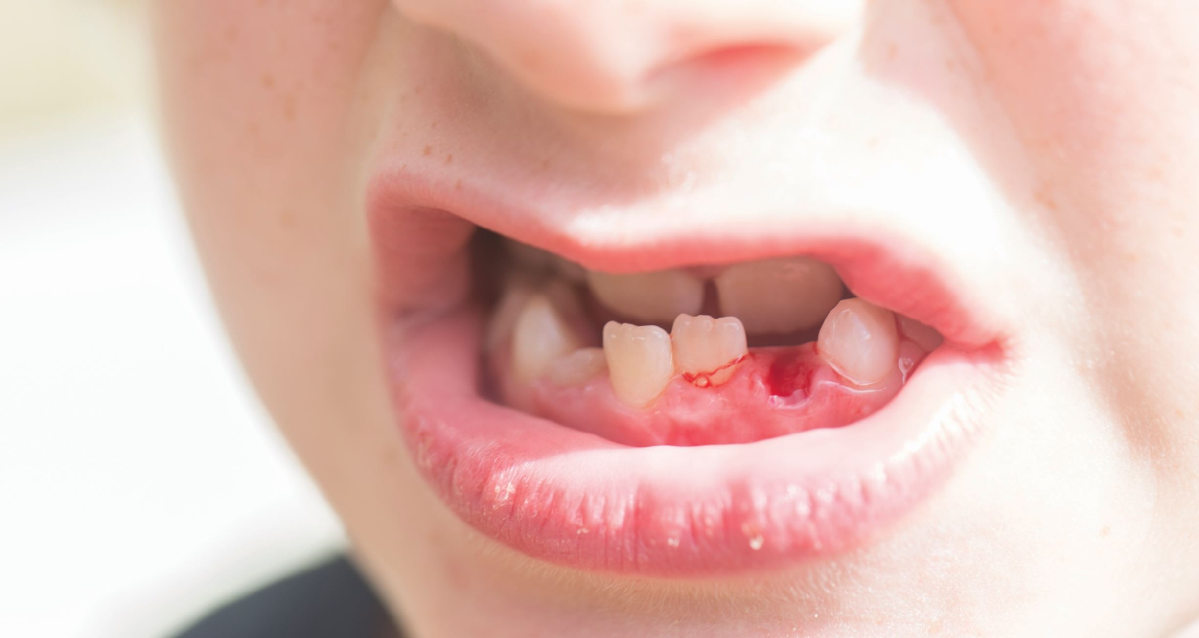
Dental implants are a predictable and widely used solution for tooth replacement. But did you know that even a small mistake in the planning or the care phase can predispose the implant to peri-implantitis, one of the most challenging complications.
Stats reveal that nearly 23% of implants are at risk of failure due to peri-implantitis¹. The good news is that peri-implantitis is preventable. A few preventive protocols in place can ensure long-term success of implants.
Here are 5 simple yet effective steps to prevent peri-implantitis:
1. Preoperative Assessment and Planning
The first prevention step begins even before the implant placement. The in-depth analysis of the patient’s oral and systemic health is essential. Clinicians should:
- Take a note of existing periodontal status and manage active infections2
- Bone quality and quantity both need to be in good condition for implant placement.
- Help patients understand the importance of oral hygiene in maintaining implant health3.
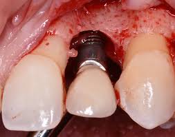
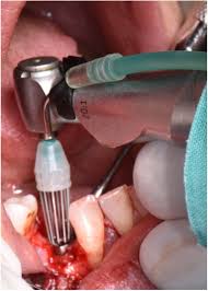
2. Aseptic Surgical Protocols
The surgical phase is the foundation of the chairside prevention strategy. Strict asepsis measures minimize the risk of bacterial contamination, which can predispose implants to inflammation. Important steps include:
- Using sterile instruments and components.
- Avoiding overheating during osteotomy and controlling flap trauma.
- Ensuring precise implant positioning for adequate soft tissue coverage.
Following these protocols reduces the risk of bacterial growth, which in turn supports long term peri-implant tissue health.
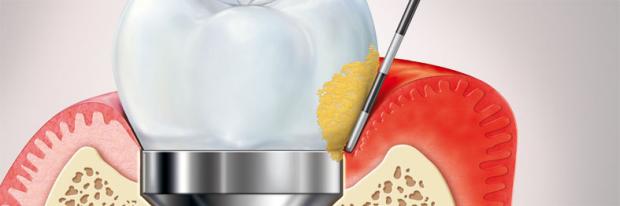
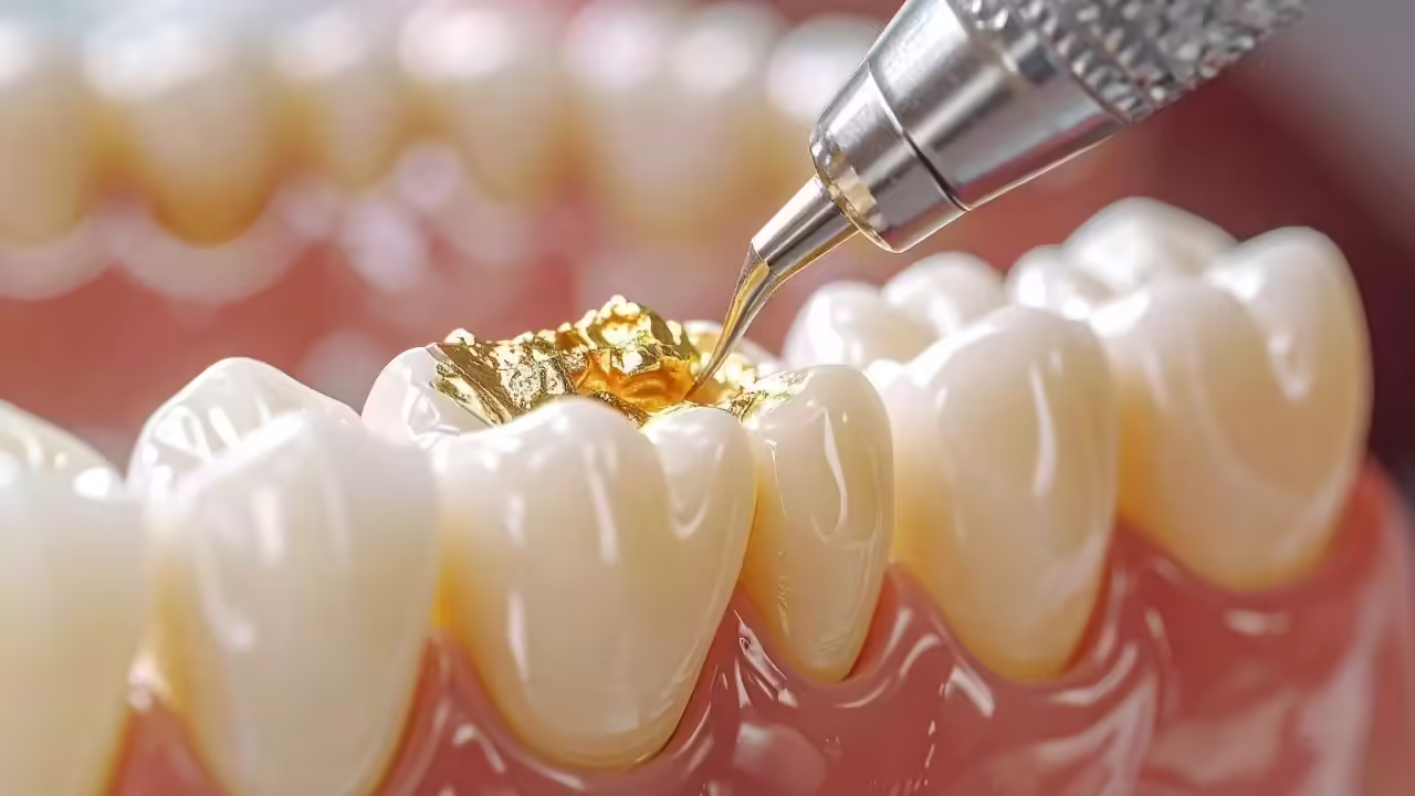
3. Prosthetic Phase Protocols
After implant placement, dentists should focus on restorative protocols. Making sure this phase is correct can significantly impact peri-implant health3. This includes:
- Remove excess cement carefully after crown placement, as residual cement is known to cause peri-implant inflammation3.
- Ensure proper occlusion as improper occlusion can cause overloading that may compromise bone support.
- Use precision impression techniques to prevent tissue trauma during the prosthetic procedure.
Strict adherence to these protocols restricts bacterial growth.
4. Regular Monitoring and Hygiene Reinforcement
Continuous monitoring and maintaining hygiene are the key pillars of peri-implantitis prevention. It includes:
- Use implant-safe instruments, like plastic or titanium scalers, for cleaning.
- Perform routine probing around implants to detect early signs of inflammation.
- Glycine powder air polishing to reduce plaque and reduce peri-implant inflammation4.
- Erythritol powder air polishing to remove biofilm from rough implant surfaces4.
- Educate patients about effective home care, including interdental brushes and microbial rinses.
Regular monitoring is the best way to aid early detection and reduce the risk of infection.
5. Adjunctive Chairside Measures
These chairside measures can further lower the risk of peri-implantitis:
- Apply antimicrobial gels or rinses during maintenance visits.
- Consider local antibiotic delivery for high-risk cases.
- Use laser-assisted debridement for biofilm control in a minimally invasive manner.
These additional routine chairside protocols provide better preventive outcomes.
Final Takeaway
By focusing on these few additional pointers, dentists can prevent peri-implantitis.
These are evidence-backed strategies not only ensure implant functionality and longevity but also enhance patient satisfaction and trust.
|
Phase |
Key Protocols/Actions |
Purpose/Tip |
|
1. Preoperative Assessment |
Periodontal evaluation, systemic health review, bone assessment, patient education |
Identify risk factors, treat active disease, and prepare patient for optimal implant outcomes |
|
2. Surgical Phase |
Strict asepsis, minimize flap trauma, controlled osteotomy, accurate implant positioning |
Reduce bacterial contamination, preserve soft tissue, ensure proper bone support |
|
3. Prosthetic Phase |
Remove excess cement, check occlusion, precision impressions, polish restoration margins |
Prevent localized inflammation, avoid overload, minimize tissue trauma, reduce plaque retention |
|
4. Maintenance & Monitoring |
Peri-implant probing, professional cleaning, air-polishing with glycine powder, reinforce home care |
Detect early inflammation, remove biofilm safely, ensure patient compliance |
|
5. Chairside Adjuncts |
Antimicrobial gels/rinses, local antibiotic delivery, laser-assisted debridement |
Enhance bacterial control, manage high-risk cases, improve biofilm removal effectiveness |
References:
- Takefuji Y. Dental implant prevalence and durability: A concise review of factors influencing success and failure. Biomaterials and Biosystems. 2025 Feb 15:100109.
- Hong I, Koo KT, Oh SY, Park HW, Sanz-Martín I, Cha JK. Comprehensive treatment protocol for peri-implantitis: an up-to date narrative review of the literature. Journal of periodontal & implant science. 2024 Jan 4;54(5):295.
- AlJasser RN, AlSarhan MA, Alotaibi DH, AlOraini S, Ansari AS, Habib SR, Zafar MS. Analysis of prosthetic factors affecting peri-implant health: an in vivo retrospective study. Journal of Multidisciplinary Healthcare. 2021 May 25:1183-91.
- Baldi D, De Giorgis L, Menini M, Motta F, Colombo J. Efficacy of instruments for professional oral hygiene on dental implants: A systematic review. Applied Sciences. 2021 Dec 21;12(1):26.
Related Contents
Video
Management of Traumatic Tooth Avulsion: When & How?
Traumatic tooth avulsion is a true dental emergency requiring immediate and structured management to...
Article
Prosthetic Design Factors That Prevent Peri-Implant Bone Loss
You placed the implant perfectly. Osseointegration was flawless. But three years later, there is 3mm...
Article
Treatment Algorithms for Peri-Implant Mucositis and Peri-Implantitis
You can prevent most peri-implant disease. But once it develops, treatment becomes less predictable....
Article
Implant Surface Decontamination: What Actually Works
Treating peri-implantitis starts with one critical step, which is decontaminating the implant surfac...
Article
Growth & Facial Asymmetry: When to Worry
Only a few dentists can notice it and realize the asymmetry can signal an underlying skeletal imbala...
Article
Orthodontic Red Flags Every Dentist Should Recognize: Functional Habits and Airway Cues
Some malocclusions cases stall for reasons you can’t see on a scan, as not...
Article
Tooth Eruption & Space Management: What Orthodontists Should Watch For
Tooth eruption may not alwaxfys fit the predictable biological timeline. A delayed or ecto...
Article
Communication & Referral Timing: Getting It Right
You have identified a problem, maybe a severe Class III malocclusion in a 9-year-old, or it is an im...