
Article
Impression Methods That Guarantee Better-Fitting CPDs
Better-fitting cast partial dentures (CPDs) begin with accurate impressions. The impression phase is not just a mechanical step—it is the foundation for retention, stability, and patient comfort. Recent research emphasizes that both conventional and digital methods have unique strengths, and mastering them ensures predictable outcomes for practicing dentists.
Why Impressions Matter
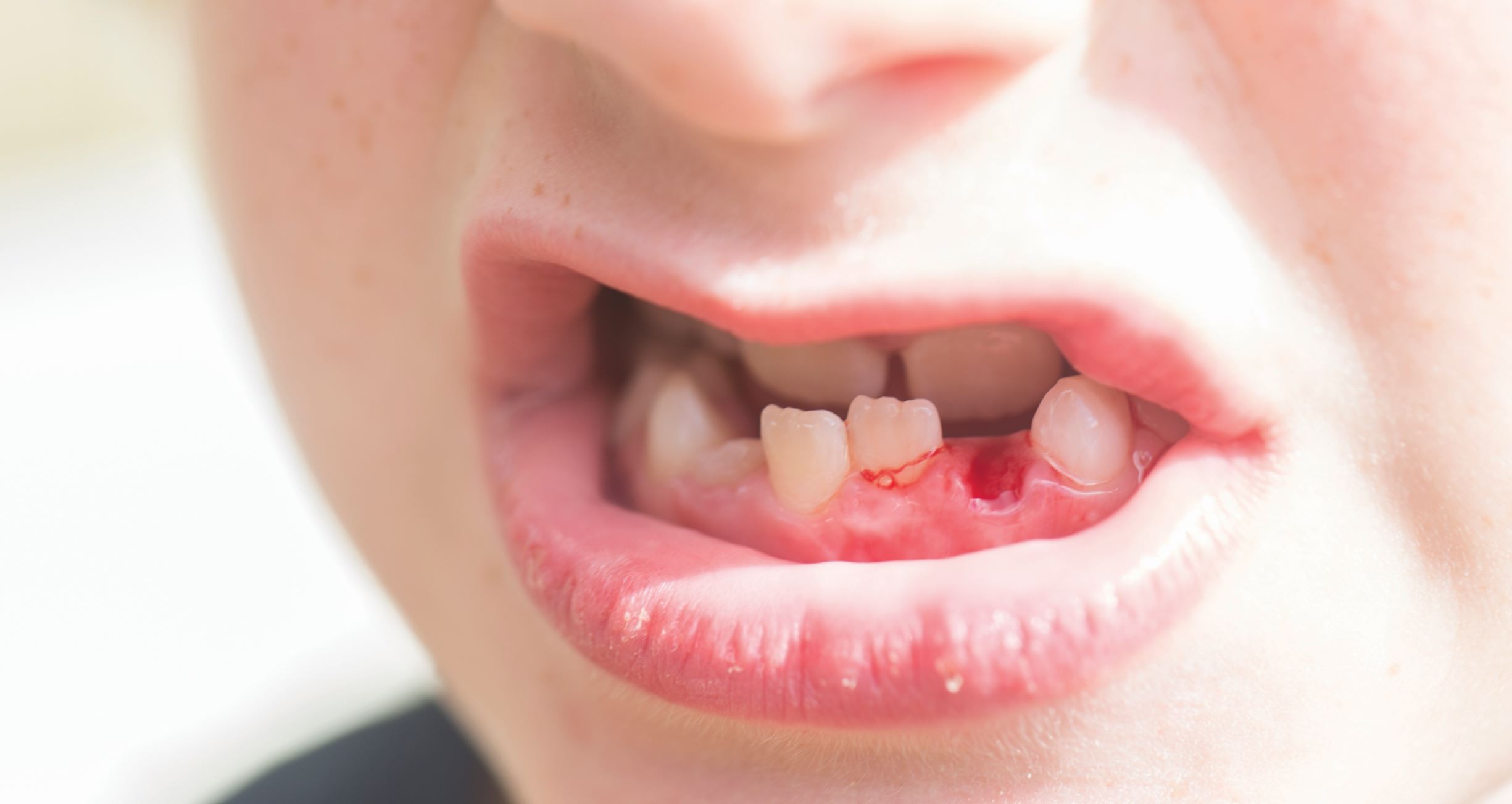
An accurate impression records the teeth, edentulous ridges, and soft-tissue contours essential for a stable removable partial denture. Because the mucosa in free-end saddles can displace up to 1.3 mm, far more than the 0.1 mm movement of an abutment tooth, any inaccuracy at this stage can lead to tissue overload, abutment stress, reduced retention, and patient discomfort1.
Since the master cast, surveying, and final design depend entirely on this initial anatomical record, a precise impression forms the foundation for a well-fitting and long-lasting prosthesis.
Conventional Impression Techniques
- Selective-Pressure Technique: This approach records the primary stress-bearing areas under controlled loading while relieving non-stress-bearing tissues to minimise tissue displacement and ensure framework support.
- Altered-Cast Technique: The altered cast technique improves the fit and function of distal-extension prostheses by achieving better tooth-tissue adaptation. It helps distribute forces more evenly, reduces stress on abutment teeth, preserves the residual ridge, and minimises denture base movement. Evidence also shows improved tissue health, comfort, and long-term stability2.
- Border-Moulding and Functional Impressions: Border-moulding involves shaping the tray borders by manipulating adjacent soft tissues to reproduce vestibular contours and establish proper extension, usually using low-fusing compound or silicone putty.
In distal-extension cases, functional impressions capture the mucosa under controlled loading, acknowledging its greater displacement compared with teeth. This minimizes excessive tissue compression, reduces stress on abutment teeth, and promotes more favourable force distribution1.
Digital Impression Methods3
- Digital impressions are the first step of the CAD-CAM workflow, captured through:
- Direct intraoral scanning, or
- Indirect extraoral scanning of a stone cast.
- A fully digital workflow uses an intraoral scanner to obtain the definitive RS-STL scan without any physical impression.
- A combined analog–digital workflow begins with a conventional impression and cast, which is then digitized using a laboratory scanner.
- Digital scans of partially dentate arches can achieve accuracy comparable to conventional impressions.
- Capturing edentulous soft-tissue areas is more challenging due to tissue compressibility and shape changes during scanning.
- Fully digital workflows can provide precise framework adaptation when the impressions are captured accurately.
Putting It All Together
With both conventional and digital techniques offering distinct advantages, clinicians must understand when and how to apply each approach. Translating these methods into everyday practice requires precision, proper technique selection, and attention to detail. The following pro tips help ensure predictable, high-quality impressions.
Pro Tips for Better Impressions
- Always dry and isolate the field before recording impressions to avoid distortion4.
- Use custom trays for partially edentulous arches to ensure uniform thickness of impression material.
- For distal extension cases, consider the altered cast technique to improve tissue support2.
- In digital workflows, scan in segments (quadrants) for better accuracy, especially in long-span arches.
- Communicate clearly with the lab—note areas of concern (mobile tissue, deep undercuts) so they can plan accordingly.
Things to Avoid
- Avoid overextension of borders, which can cause sore spots and instability.
- Do not rely solely on stock trays for complex cases—these often miss critical landmarks.
- Avoid excessive pressure during functional impressions, which may distort soft tissue contours.
- In digital impressions, avoid rapid scanning movements—they can create stitching errors and inaccurate data.
- Never skip verification of the impression/scan before sending to the lab; small errors magnify in the final framework.
Clinical Benefits for CPDs
By integrating modern impression methods and following best practices, dentists can expect:
- Improved fit and retention
- Reduced adjustments and remakes
- Enhanced patient comfort
- Better lab communication
Final Takeaway
Impression methods—whether conventional or digital—are the cornerstone of CPD success. For practicing dentists, combining traditional functional techniques with modern digital workflows, while following pro tips and avoiding common pitfalls, guarantees better-fitting CPDs and happier patients.
Reference
- de Moraes Melo Neto CL, de Oliveira Costa F, Reginato VF, Neves ACC, de Sousa Menezes M, de Almeida EO. Removable partial denture – functional impression techniques: Review. Prague Med Rep. 2023;124(4):380–393.
- Rashmi M, Nagi D, Kiruthiga D, Soneya Punith, Senbagavalli S. Altered cast technique: A case report. Int J Dent Sci. 2025;7(2):44-46.
- Elgamal M, Ibrahim AM, Fadl BT, Ragheb NA. Accuracy assessment of removable partial denture frameworks fabricated by selective laser melting using two different workflows: A cross-over clinical study. BMC Oral Health. 2025 May 28;25(1):824.
- Parameshwari G, Chittaranjan B, Sudhir N, Anulekha-Avinash CK, Taruna M, Ramureddy M. Evaluation of accuracy of various impression techniques and impression materials in recording multiple implants placed unilaterally in a partially edentulous mandible-An in vitro study. Journal of clinical and experimental dentistry. 2018 Apr 1;10(4):e388.
Related Contents
Video
Management of Traumatic Tooth Avulsion: When & How?
Traumatic tooth avulsion is a true dental emergency requiring immediate and structured management to...
Article
Prosthetic Design Factors That Prevent Peri-Implant Bone Loss
You placed the implant perfectly. Osseointegration was flawless. But three years later, there is 3mm...
Article
Treatment Algorithms for Peri-Implant Mucositis and Peri-Implantitis
You can prevent most peri-implant disease. But once it develops, treatment becomes less predictable....
Article
Implant Surface Decontamination: What Actually Works
Treating peri-implantitis starts with one critical step, which is decontaminating the implant surfac...
Article
Growth & Facial Asymmetry: When to Worry
Only a few dentists can notice it and realize the asymmetry can signal an underlying skeletal imbala...
Article
Orthodontic Red Flags Every Dentist Should Recognize: Functional Habits and Airway Cues
Some malocclusions cases stall for reasons you can’t see on a scan, as not...
Article
Tooth Eruption & Space Management: What Orthodontists Should Watch For
Tooth eruption may not alwaxfys fit the predictable biological timeline. A delayed or ecto...
Article
Communication & Referral Timing: Getting It Right
You have identified a problem, maybe a severe Class III malocclusion in a 9-year-old, or it is an im...