
Article
Early Enamel Demineralization After Bracket Placement: What the Evidence Shows
Fixed orthodontic appliances improve alignment but also create a plaque-retentive environment that makes cleaning more challenging. Brackets and bands interfere with natural self-cleansing and promote areas of stagnation, increasing the likelihood of early enamel demineralization during treatment.
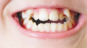
Clinical evidence shows that white spot lesions (WSLs) can appear as early as the fourth week after bonding, especially in patients with poor oral hygiene. Recent studies report a wide WSL prevalence range of 33.8% to 97%, reflecting differences in assessment methods and patient-related factors1. These findings highlight the rapid onset and high risk of WSLs, emphasizing the need for early and consistent preventive strategies.
What Actually Changes?
Multiple laboratory and clinical studies report a rapid shift in plaque composition after appliance placement, with increases in acidogenic species such as Streptococcus mutans and greater biofilm biomass adjacent to bracket bases. These microorganisms metabolize dietary sugars into acids that lower local pH and begin the diffusion‑driven loss of mineral from the enamel subsurface — the microscopic start of a WSL2.
How Common is It?
WSLs are one of the most frequent complications of fixed-appliance therapy.
- A 2024 meta-analysis of 57 studies (≈9,100 patients) reported a 55% prevalence and 34% incidence of new lesions during treatment3. Earlier pooled data show even higher rates, with incidence around 46% and prevalence up to 68%4.
- Clinical studies mirror these findings. One cohort showed WSLs rising from 11.9% at baseline to 37.3% at 6 months and 46.6% at 12 months5, while another reported that 78.6% of patients developed at least one new lesion by the end of treatment6.
- Across evidence, roughly one in two orthodontic patients develop WSLs, with variability driven by diagnostic methods, baseline enamel status, treatment duration, and oral-hygiene compliance.
What Does This Mean in Practice?
- First, expect the greatest risk during the early months after bonding. Intensify prevention and surveillance during that period rather than waiting for lesions to become obvious at mid-treatment or debonding.
- Second, diagnostic sensitivity matters: adjunctive tools such as fluorescence-aided inspection7 or standardized photography identify more early lesions than unaided visual exam and let you act while remineralization is still feasible.
Device and Material Factors Matter
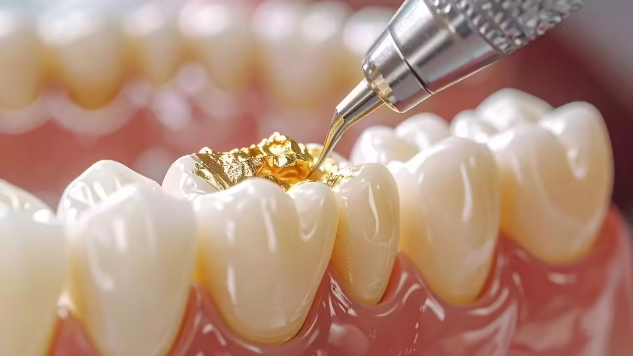
Bracket material, adhesive choice, and coatings can influence enamel demineralization by affecting plaque retention and bacterial adhesion. Fluoride-releasing adhesives (e.g., RMGIC, GIC) and bioactive adhesives containing calcium or phosphate particles have been shown to reduce white spot lesions compared with conventional resins8,9. Applying protective sealants around bracket bases can further minimize risk, making material selection a key part of evidence-based prevention for high-risk patients.
Final Takeaways for the Practicing Dentist
- Treat bonding as a high-risk event; assess caries risk beforehand.
- Document baseline enamel with photos.
- Give focused hygiene instructions tailored to the patient.
- Schedule early follow-ups in the first 4–8 weeks.
- Start non-invasive remineralization at first signs of demineralization.
- Prevention and early detection preserve enamel and reduce future restorative needs.
Reference
- Hamdi K, Elsebaai A, Abdelshafi MA, Hamama HH. Remineralization and anti-demineralization effect of orthodontic adhesives on enamel surrounding orthodontic brackets: a systematic review of in vitro studies. BMC Oral Health. 2024 Nov 28;24(1):1446.
- Al-Blaihed D, El Meligy O, Baghlaf K, Aljawi RA, Abudawood S, El Meligy OA. White spot lesions in fixed orthodontics: a literature review on etiology, prevention, and treatment. Cureus. 2024 Jul 29;16(7).
- Hussain U, Wahab A, Kamran MA, Alnazeh AA, Almoammar S, Alshahrani SS, Niazi FH, Alam S, Arif N, Campobasso A, Pandis N. Prevalence, Incidence and Risk Factors of White Spot Lesions Associated With Orthodontic Treatment–A Systematic Review and Meta‐Analysis. Orthodontics & Craniofacial Research. 2025 Apr;28(2):379-99.
- Sundararaj D, Venkatachalapathy S, Tandon A, Pereira A. Critical evaluation of incidence and prevalence of white spot lesions during fixed orthodontic appliance treatment: A meta-analysis. Journal of International Society of Preventive and Community Dentistry. 2015 Nov 1;5(6):433-9.
- Jha AK, Chandra S, Shankar D, Murmu DC, Noorani MK, Tewari NK, Jha AK, MALAYSIA MK, kumar Tewari N. Evaluation of the prevalence of white spot lesions during fixed orthodontic treatment among patients reporting for correction of malocclusion: a prevalence study. Cureus. 2023 Jul 19;15(7).
- Gönder HY, Yıldırım M, Metli ŞN. Incidence of white spot lesions and DMFT among patients treated with comprehensive orthodontics. International Dental Research. 2022 Dec 31;12(Suppl. 1):85-9.
- BONFANTE FD, LONGHI AV, DEMARCHI M, CASTRO DM, RHODEN FK, SÓ GB, CARLI JP, CORAZZA PH, BELLO YD, STEIER L, FIGUEIREDO JA. Assessment of diagnostic accuracy of reveal® autofluorescent dental loupes for detection of biofilm, demineralization, and caries around orthodontic brackets. Dental Press Journal of Orthodontics. 2025 Nov 7;30(4):e252577.
- Mahmoud GA, Gordon PH, Pretty IA, McCabe JF, Hajeer MY, Mahmoud G, Hajeer MY. Effect of Fluoride Release on Enamel Demineralization Adjacent to Orthodontic Brackets. Cureus. 2023 Sep 28;15(9).
- El Helou M, Chakar S, Nicolas E, Estephan E, Cuisinier F, Barthélemi S. Can Orthodontic Adhesive Systems Inhibit the Formation and Development of White Spot Lesions During Fixed Orthodontic Treatment? A Systematic Review. The journal of adhesive dentistry. 2024 Oct 14;26:b5781299.
Related Contents
Video
Management of Traumatic Tooth Avulsion: When & How?
Traumatic tooth avulsion is a true dental emergency requiring immediate and structured management to...
Article
Prosthetic Design Factors That Prevent Peri-Implant Bone Loss
You placed the implant perfectly. Osseointegration was flawless. But three years later, there is 3mm...
Article
Treatment Algorithms for Peri-Implant Mucositis and Peri-Implantitis
You can prevent most peri-implant disease. But once it develops, treatment becomes less predictable....
Article
Implant Surface Decontamination: What Actually Works
Treating peri-implantitis starts with one critical step, which is decontaminating the implant surfac...
Article
Growth & Facial Asymmetry: When to Worry
Only a few dentists can notice it and realize the asymmetry can signal an underlying skeletal imbala...
Article
Orthodontic Red Flags Every Dentist Should Recognize: Functional Habits and Airway Cues
Some malocclusions cases stall for reasons you can’t see on a scan, as not...
Article
Tooth Eruption & Space Management: What Orthodontists Should Watch For
Tooth eruption may not alwaxfys fit the predictable biological timeline. A delayed or ecto...
Article
Communication & Referral Timing: Getting It Right
You have identified a problem, maybe a severe Class III malocclusion in a 9-year-old, or it is an im...