
Article
Pulp Vitality Monitoring: When to Intervene Endodontically?
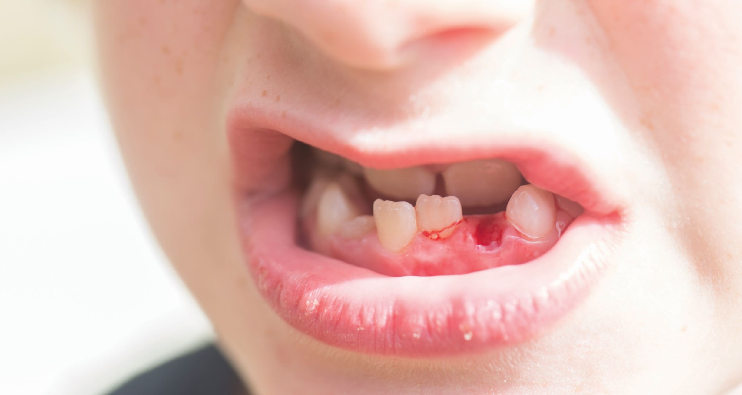
Every discolored or symptomatic tooth does not necessarily need a root canal. Modern endodontics emphasizes pulp preservation and intervening only if the tooth is non-vital or irreversible inflammation sets in1.
Since pulp necrosis can occur months or even years after trauma, regular pulp vitality monitoring is essential.
Step 1: Know About the Pulpal Spectrum
The pulp doesn’t just flip from “alive” to “dead.” It passes through a continuum, and understanding all the stages is equally important2:
|
Clinical State |
Symptoms |
Recommended Action |
|
Normal pulp |
No lingering pain, normal response to tests |
Routine monitoring |
|
Reversible pulpitis |
Sharp, transient pain; triggered by stimuli |
Remove irritant, restore tooth |
|
Irreversible pulpitis |
Spontaneous, lingering pain |
Endodontic intervention indicated |
|
Pulp necrosis |
No response to vitality tests |
Root canal or extraction |
Always correlate pulp-test results with clinical findings rather than relying on any single test.
Traumatic dental injuries disrupt the neurovascular bundle entering the apex. Teeth with open apices have significantly better revascularization potential than mature teeth3,4.
Mature teeth have higher rates of pulp necrosis across all luxation types, especially if there are complicated injuries.
Step 2: Use the Right Vitality Tests
Find out about different tests and how to decide the one most suitable for you -
1. Cold Test - Still the Gold Standard
- Use refrigerant spray on the cotton pellet.
- A quick, sharp response indicates vital pulp.
- Delayed or no response indicates compromised pulp.
- Preferred choice of pulp testing in routine and trauma follow-ups5.
2. Electric Pulp Test (EPT)
- Evaluates neural response of the tooth and does not assess vascular flow.
- False negatives possible in recently traumatized or calcified teeth6.
- Use as adjunct, not standalone.
3. Laser Doppler Flowmetry & Pulse Oximetry
- Measure blood flow in the pulp, which is ideally a true vitality indicator2,7.
- Useful for immature teeth or trauma cases where EPT can be unreliable.
- Compact, wireless models available now making the use easier.
4. Thermal (Heat) & Test Cavities
- Heat helps identify irreversible pulpitis that may present as a lingering, dull ache.
- Test cavity without anesthesia is last resort
Chairside Tip: Always test adjacent and contralateral teeth to avoid false interpretations2.
Step 3: Timing the Intervention2
|
Scenario |
Key Findings |
Action |
|
Deep carious lesion, sharp transient pain |
Short, stimulus-linked pain |
Remove decay, indirect pulp capping |
|
Lingering pain (>30 s) or spontaneous throbbing |
Consistent with irreversible pulpitis |
Start RCT |
|
No cold/EPT response + discoloration |
Suggestive of necrosis |
Radiograph + confirm with another test |
|
Recently traumatized tooth |
Temporary loss of response possible for ≤ 12 weeks |
Monitor with vitality testing every 3–4 weeks |
|
Partial pulp exposure in young permanent tooth |
Bleeding, but controllable |
Vital pulp therapy (MTA/Biodentine) |
Step 4: Radiographic Correlation
Radiographic interpretation can be related to pulp testing as it may reveal early structural changes that reflect loss of vitality or pulpal inflammation.
A widened periodontal ligament (PDL) space is usually an early sign of inflammation, which can be seen before clinical symptoms appear.
Loss of lamina dura or building periapical radiolucency can indicate transition from reversible pulpitis to irreversible pulpitis or apical periodontitis.
In advanced cases, internal or external root resorption may become visible that suggests pulpal necrosis or chronic inflammation.
Radiographic assessment along with tests such as cold, electric pulp testing, or vitality assessment can help in confirming pulpal status, and guides timely endodontic intervention2.
Step 5: Documentation & Review
While we keep talking about ideal pulp testing, it is equally important to understand the value of documentation.
- Always record baseline test results, stimulus type, and patient responses.
- Re-evaluate at every recall, especially after trauma or deep restorations.
- Vitality tests help decide whether to continue monitoring or intervene.
In post-trauma or revascularization cases, continued pulp testing helps make long-term success decisions.
Final Takeaway
A systematic, evidence-based vitality-monitoring protocol prevents unnecessary RCTs and helps pulp survival.
You can always combine clinical judgment + modern diagnostics + periodic reassessment for predictable outcomes.
References
- Lima TFR, Dos Santos SL, Fidalgo TKS, Nogueira Leal ES. Vitality tests for pulp diagnosis of traumatized teeth: a systematic review. J Endod. 2019;45(5):490-499.
- Duncan HF, Kirkevang LL, Peters OA, El-Karim I, Krastl G, Del Fabbro M, et al. Treatment of pulpal and apical disease: the European Society of Endodontology (ESE) S3-level clinical practice guideline. Int Endod J. 2023;56(S3):238-295.
- Levin L, Day PF, Hicks L, O'Connell A, Fouad AF, Bourguignon C, et al. International Association of Dental Traumatology guidelines for the management of traumatic dental injuries: General introduction. Dent Traumatol. 2020;36(4):309-13.
- Bourguignon C, Cohenca N, Lauridsen E, Flores MT, O'Connell AC, Day PF, et al. International Association of Dental Traumatology guidelines for the management of traumatic dental injuries: 1. Fractures and luxations. Dent Traumatol. 2020;36(4):314-30
- Mainkar A, Kim SG. Diagnostic accuracy of 5 dental pulp tests: a systematic review and meta-analysis. J Endod. 2018;44(5):694–702.
- Çağırır Dindaroğlu F, Özay Güngör N. Comparison of the vitality test with sensitivity tests in mature and immature teeth: clinical trial. BMC Oral Health. 2024;24(1):613.
- Bux M, Adam M. Accuracy of vitality and sensibility testing in mature and immature anterior teeth: a clinical trial. Endodontics. 2024 Aug 20.
Related Contents
Video
Management of Traumatic Tooth Avulsion: When & How?
Traumatic tooth avulsion is a true dental emergency requiring immediate and structured management to...
Article
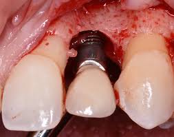
Prosthetic Design Factors That Prevent Peri-Implant Bone Loss
You placed the implant perfectly. Osseointegration was flawless. But three years later, there is 3mm...
Article
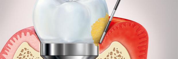
Treatment Algorithms for Peri-Implant Mucositis and Peri-Implantitis
You can prevent most peri-implant disease. But once it develops, treatment becomes less predictable....
Article
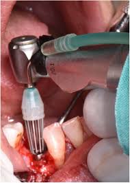
Implant Surface Decontamination: What Actually Works
Treating peri-implantitis starts with one critical step, which is decontaminating the implant surfac...
Article
Growth & Facial Asymmetry: When to Worry
Only a few dentists can notice it and realize the asymmetry can signal an underlying skeletal imbala...
Article
Orthodontic Red Flags Every Dentist Should Recognize: Functional Habits and Airway Cues
Some malocclusions cases stall for reasons you can’t see on a scan, as not...
Article
Tooth Eruption & Space Management: What Orthodontists Should Watch For
Tooth eruption may not alwaxfys fit the predictable biological timeline. A delayed or ecto...
Article
Communication & Referral Timing: Getting It Right
You have identified a problem, maybe a severe Class III malocclusion in a 9-year-old, or it is an im...