Article
Oral Lesions Every Dentist Must Detect Early: Recognizing the Silent and Functional Signs
Oral lesions signal an underlying health issue. It could range from minor irritations to serious conditions such as oral cancers.
Although the early signs are easily overlooked as they are mostly painless, timely recognition could influence the prognosis significantly.
For instance, oral cancers when diagnosed early at a localized stage have an 88% five-year survival rate in comparison to advanced stage whose survival rate decreases to 36.9%1.
Thus, recognizing both the visible signs and functional changes will equip the dentists to identify and treat the lesions quickly.
1. Leukoplakia: Persistent White Patches
Signs & Clinical Insight1:
- Homogeneous leukoplakia
- Predominantly white lesion
- Uniform, flat, thin appearance
- May exhibit shallow cracks
- Surface can be smooth, wrinkled, or corrugated
- Constant texture throughout
- Non-homogeneous leukoplakia
- Predominantly white or white-and-red lesion (erythroleukoplakia)
- May appear irregularly flat, nodular, or exophytic
- Nodular lesions
- White patches or nodules
- Present on an erythematous (red) base
- Exophytic lesions
- Irregular blunt or sharp projections
Functional Sign:
- Usually none, occasionally mild discomfort during mastication if on the buccal mucosa2
Pro-Tip: Roll lesions with gauze, use magnification, and document with intraoral photographs. Persistent lesions beyond two weeks require biopsy4.
2. Erythroplakia: High-Risk Red Lesion5
Signs & Clinical Insight:
- Red, velvety patches, frequently asymptomatic
- High malignant potential (dysplasia/carcinoma)6
Functional Sign:
- Rarely, slight discomfort on swallowing if on the floor of the mouth
Pro-Tip: Inspect with loupes, palpate for induration, biopsy immediately7.
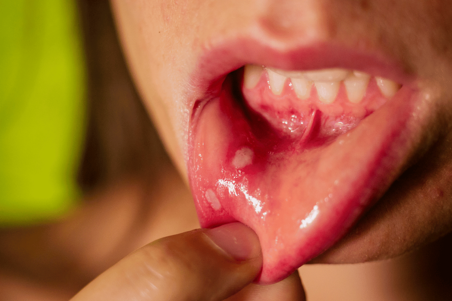
3. Oral Lichen Planus (OLP)8,9
Signs & Clinical Insight:
- Chronic inflammatory disease affecting oral mucosa; ~1.5% prevalence, more common in females
- May occur alone or with other mucosal/skin involvement
- Six main types: reticular, erosive/ulcerative (including bullous), papular, plaque-like, erythematous, atrophic
- Reticular type often asymptomatic; erosive type causes burning pain, triggered by sour/spicy foods
- Erosive subtype has ~1% risk of malignant transformation over 3–6 years
Functional Signs:
- Burning sensation or sensitivity to acidic/spicy foods
- Rare difficulty in mastication if erosive lesions are extensive
Pro-Tip:
- Combine clinical exam with histopathology; biopsy representative lesions
- Review medications to rule out lichenoid drug reactions
- Use serial photographs to monitor progression
- Biopsy persistent or suspicious changes early
4. Early Oral Cancer (OSCC)10
Signs & Clinical Insight:
- Non-healing ulcers, small nodules and mucosal thickening
- Mostly at the early stage it is painless
Functional Sign:
- Early dysphagia, difficulty in chewing and speech may change.
- If lateral border is involved, then restriction in tongue mobility could be observed slightly.
Pro-Tip: Do inspection and palpation. Document induration and opt for biopsy if lesion persists.
5. Actinic Cheilitis11
Signs & Clinical Insight:
- Excessive UV exposure causes this chronic and malignant lips disorder.
- Blurred demarcation between lip vermilion and skin.
- Erythema.
- Scaling, and dryness of lower lip.
- In advanced cases, plaques, crusts or ulcerations may appear.
- Unfortunately, as the evolution of the lesion is slow and often asymptomatic, it could be easily mistaken for aging.
- If untreated, it could be become malignant, and the transformation rate is found to be ~10–30%.
- Smoking, alcohol, light skin, outdoor exposure, and limited use of lip sunscreen increases the risk of development.
Functional Signs:
- Asymptomatic initially.
- In the advanced stage, concerns associated with lip mobility, eating, or speech may be observed.
Pro-Tip:
- Monitor for incessant dryness, scaling, or plaques.
- Do biopsy of suspicious or non-healing lesions for assessing the epithelial dysplasia.
6. Submucous Fibrosis12
Signs & Clinical Insight:
- Blanched and dense bands of fibrotic mucosa.
Functional Sign:
- Reduced mouth opening, speech limitations and difficulty with mastication.
Pro-Tip: Measure the opening of the mouth, palpate fibrotic bands and monitor the progression.
7. Papillary Lesions (Papilloma, Verrucous Carcinoma)13
Signs & Clinical Insight:
- Small and painless exophytic growths.
Functional Sign:
- Usually minimal but large lesions may interfere with phonation or mastication.
Pro-Tip:
- Inspect surface texture.
- Monitor the growth.
- Do biopsy if lesions persist.
8. Pigmented Lesions / Melanotic Macules14
Signs & Clinical Insight:
- Asymptomatic dark oral patches.
Functional Sign:
- Unless extensive, it does not impact function.
- Pro-Tip: Document with photos, do biopsy if lesion keeps evolving.
Enhancing Detection: Diagnostic Adjuncts
Clinical inspection coupled with adjunctive tools can improve the accuracy of diagnosis15. Some of these tools are:
- Autofluorescence devices: They aid by highlighting the suspicious mucosa
- Brush biopsies: It helps in cytological evaluation of ambiguous lesions
- Toluidine blue staining: It helps in the recognition of high-risk areas
Final Takeaway
Early recognition of lesion’s silent and functional signs can help to detect malignant oral lesions. Timely intervention can be provided by integrating inspection, palpation, patient questioning and photographic documentation. Along with this, judicious use of adjunctive diagnostics will act as icing on the cake in improving recovery rates.
References
- American Cancer Society. Survival Rates for Oral Cavity and Oropharyngeal Cancer. American Cancer Society; 2025 Jun 26. Available from: https://www.cancer.org/cancer/types/oral-cavity-and-oropharyngeal-cancer/detection-diagnosis-staging/survival-rates.html
- Waghmare PV, Raut DL. OROPHARYNGEAL NON SCRAPABLE WHITE LESIONS-A DIAGNOSTIC DILEMMA. Journal of Interdisciplinary Dental Sciences. 2018 Jan;7(1).
- Bankvall M, Dabelsteen E, Holmstrup P, Johannessen AC, Jontell M, Neppelberg E, Rautava J. Common oral mucosal lesions. Den norske tannlegeforenings Tidende. 2024 Feb 15;134(2):126-38.
- Mortazavi H, Safi Y, Baharvand M, Jafari S, Anbari F, Rahmani S. Oral white lesions: an updated clinical diagnostic decision tree. Dentistry journal. 2019 Feb 7;7(1):15.
- Öhman J, Zlotogorski-Hurvitz A, Dobriyan A, Reiter S, Vered M, Willberg J, Lajolo C, Siponen M. Oral erythroplakia and oral erythroplakia-like oral squamous cell carcinoma–what’s the difference?. BMC Oral Health. 2023 Nov 13;23(1):859.
- Lorenzo‐Pouso AI, Lafuente‐Ibáñez de Mendoza I, Pérez‐Sayáns M, Pérez‐Jardón A, Chamorro‐Petronacci CM, Blanco‐Carrión A, Aguirre‐Urízar JM. Critical update, systematic review, and meta‐analysis of oral erythroplakia as an oral potentially malignant disorder. Journal of Oral Pathology & Medicine. 2022 Aug;51(7):585-93.
- Öhman J, Zlotogorski-Hurvitz A, Dobriyan A, Reiter S, Vered M, Willberg J, Lajolo C, Siponen M. Oral erythroplakia and oral erythroplakia-like oral squamous cell carcinoma–what’s the difference?. BMC Oral Health. 2023 Nov 13;23(1):859.
- Cramer N, Kromer D, Bootsveld JM, Sennhenn‐Kirchner S, Gerdes S, Sondermann W, Assaf K, Goebeler M, Wilsmann‐Theis D, Günther C, Kromer C. History, clinical presentation, therapy, and patient reported outcomes of mucosal lichen planus: a cross‐sectional study. JDDG: Journal der Deutschen Dermatologischen Gesellschaft. 2025 Apr;23(4):449-62.
- Manchanda Y, Rathi SK, Joshi A, Das S. Oral lichen planus: an updated review of etiopathogenesis, clinical presentation, and management. Indian Dermatology Online Journal. 2024 Jan 1;15(1):8-23.
- Adamson OO, Erinoso O, Oluwakuyide R, Amao A, Effiom O, Gbotolorun OM. Association between non-healing precancerous oral lesions and ulcers with tobacco smoking: A population-based study. Oral Oncology Reports. 2024 Jun 1;10:100428.
- Carneiro MC, Quenta-Huayhua MG, Peralta-Mamani M, Honório HM, Santos PS, Rubira-Bullen IR, Rubira CM. Clinicopathological analysis of actinic cheilitis: a systematic review with meta-analyses. Head and Neck Pathology. 2023 Sep;17(3):708-21.
- Tang J, Liu J, Zhou Z, Cui X, Tu H, Jia J, Chen B, Dai X, Liu O. Oral submucous fibrosis: pathogenesis and therapeutic approaches. International Journal of Oral Science. 2025 Feb 1;17(1):8.
- Modak B, Kulkarni S. Clinicopathological analysis of potentially malignant and malignant verrucopapillary lesions of the oral cavity–A literature review. Oral Oncology Reports. 2024 Sep 1;11:100587.
- Abati S, Sandri GF, Finotello L, Polizzi E. Differential diagnosis of pigmented lesions in the oral mucosa: A clinical based overview and narrative review. Cancers. 2024 Jul 8;16(13):2487.
- Lau J, Guru O, Warnakulasuriya S, Balasubramaniam R, Frydrych A, Kujan O. Adjunctive aids for the detection of oral squamous cell carcinoma and oral potentially malignant disorders: A systematic review of systematic reviews. Japanese Dental Science Review. 2024 Dec 1;60:53-72.
Related Contents
Article
Prosthetic Design Factors That Prevent Peri-Implant Bone Loss
You placed the implant perfectly. Osseointegration was flawless. But three years later, there is 3mm...
Article
Treatment Algorithms for Peri-Implant Mucositis and Peri-Implantitis
You can prevent most peri-implant disease. But once it develops, treatment becomes less predictable....
Article
Implant Surface Decontamination: What Actually Works
Treating peri-implantitis starts with one critical step, which is decontaminating the implant surfac...
Article
Growth & Facial Asymmetry: When to Worry
Only a few dentists can notice it and realize the asymmetry can signal an underlying skeletal imbala...
Article
Orthodontic Red Flags Every Dentist Should Recognize: Functional Habits and Airway Cues
Some malocclusions cases stall for reasons you can’t see on a scan, as not...
Article
Tooth Eruption & Space Management: What Orthodontists Should Watch For
Tooth eruption may not alwaxfys fit the predictable biological timeline. A delayed or ecto...
Article
Communication & Referral Timing: Getting It Right
You have identified a problem, maybe a severe Class III malocclusion in a 9-year-old, or it is an im...
Article
Rotary File Fatigue – How to Prevent Separation?
File breakage in maxillary molars is not a rare sight, but definitely no less than a nightmare. But...