
Article
Minimally Traumatic Extraction and Ridge Preservation Strategies
Tooth removal sets off a biological domino effect. The alveolar ridge begins to remodel almost immediately, with studies showing up to 50% loss in ridge width within the first 3–6 months post-extraction1.
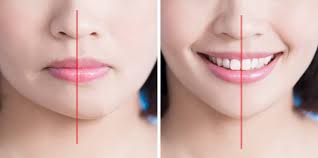
The anterior maxilla suffers the most; its buccal plate, often <1 mm thick, is prone to rapid resorption, leaving behind a flat or concave contour that compromises both function and esthetics.
Clinical Insight: Even a 1 mm buccal plate loss can mean papillary recession or a flattened gingival profile, both difficult, sometimes impossible, to restore later.
Pro Tip: Plan ridge preservation before extraction. A pre-decided protocol always outperforms an improvised one.
Extraction Redefined: From Tooth Removal to Tissue Preservation
When working in the smile zone, extraction is not a mechanical act; it’s a biological choreography. The clinician’s goal is not just to remove the tooth, but to protect the future site for an implant that will harmonize with nature.
Your Four Golden Rules
- Preserve socket walls: Avoid fracturing fragile buccal plates.
- Maintain soft-tissue architecture: Guard papillae and gingival margins like esthetic assets.
- Protect vascularity: Keep periosteum intact — it’s the lifeline of your ridge.
- Think ahead: Extraction marks the start of regeneration, not the end of surgery.
Precision in Practice: Techniques that Redefine “Atraumatic”
1. Periotome Assisted Extraction: Gentle, Controlled, Predictable
By severing periodontal fibers precisely, periotomes minimize socket trauma. Randomized trials report shorter operating times and reduced postoperative pain compared to piezotome techniques, though evidence on marginal bone preservation remains mixed2.
Best suited for single-rooted teeth where preserving socket morphology is paramount.
2. Physics Forceps: Engineering Meets Biology
A new generation of atraumatic instruments, Physics Forceps rely on a controlled rotational force rather than traditional squeezing. Systematic reviews suggest they can reduce buccal plate fractures, extraction time, and trauma, provided clinicians are well-trained3. Experts advocate including them in routine dental training for predictable results.
3. Piezosurgery: Ultrasonic Precision for Hard Tissue
When working in dense bone or with ankylosed roots, piezosurgery shines. Its ultrasonic microvibrations allow selective bone cutting, sparing soft tissue and improving precision2.
The result? A cleaner field and faster, more comfortable healing.
Choosing the Right Tool for Atraumatic Precision
Before planning ridge preservation or implant placement in the smile zone, choosing the right extraction approach is critical. The following table summarizes clinically proven techniques that help preserve bone and soft-tissue integrity while minimizing surgical trauma.
|
Technique / Tool |
Core Principle |
Best Indications |
Advantages |
|
Periotome-assisted extraction |
Gentle severing of PDL fibers |
Single-rooted teeth |
Preserves socket walls, reduces trauma |
|
Physics forceps |
Controlled rotational force |
Multi-/single-rooted teeth |
Fewer root & plate fractures |
|
Piezosurgery |
Ultrasonic selective bone cutting |
Dense bone, ankylosed roots |
Precise, less soft-tissue injury |
|
Flapless / minimal reflection |
Periosteal preservation |
Esthetic zone, thin biotype |
Maintains vascularity, ridge volume |
|
Socket inspection & immediate grafting |
Repair early, seal socket |
All esthetic extractions |
Prevents collapse, supports implant |
Table: Quick Reference Chart – Minimally Traumatic Extraction Techniques2,3,4.
Pro Tips:
- Plan before you pull: choose the least invasive tool for tooth and bone condition.
- Stay flapless whenever possible: every millimeter of periosteum preserved sustains blood flow.
- Use magnification: fine visualization reduces micro-fractures.
- Mix grafts with PRF to form sticky bone and accelerate healing.
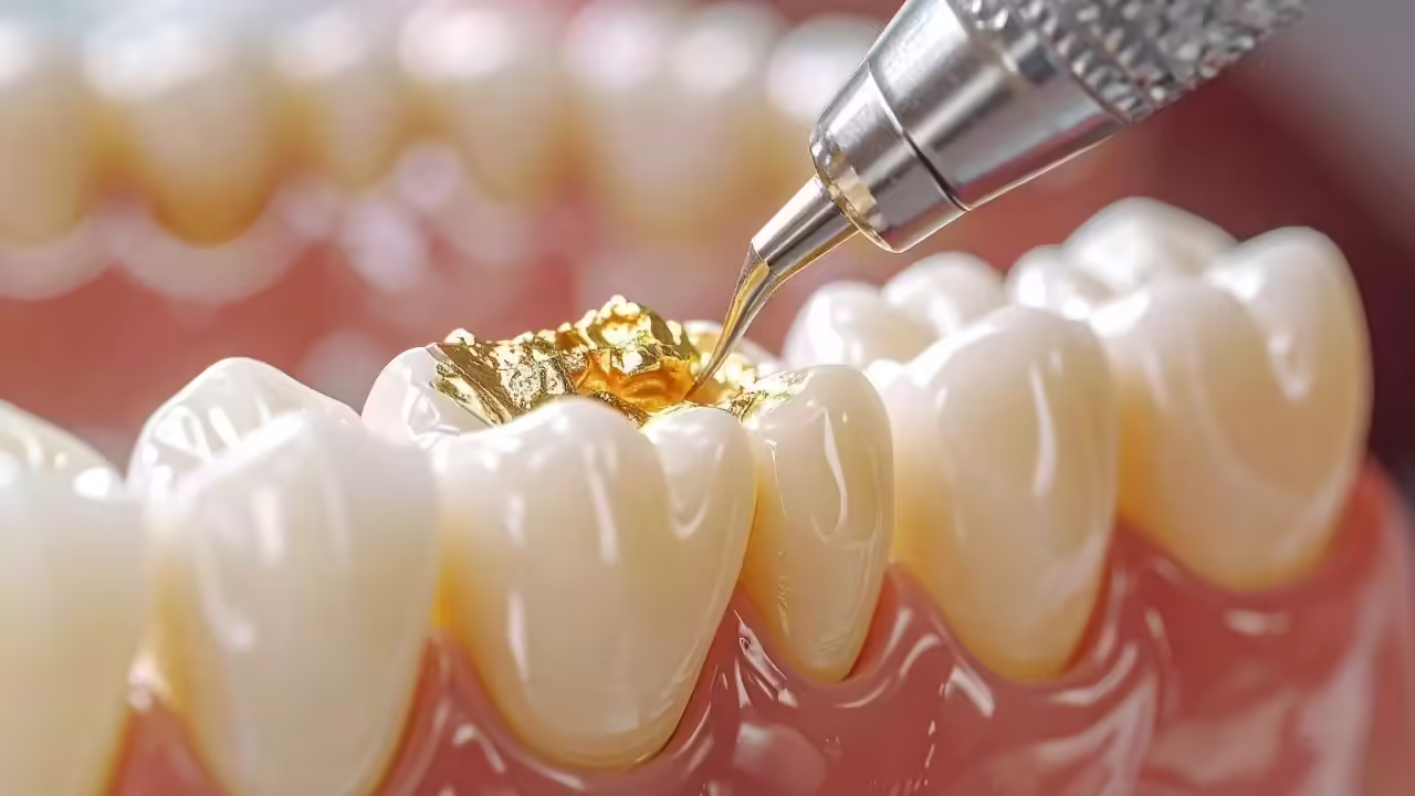
Ridge Preservation: Materials and Biologics
Post-extraction, stabilize the clot and preserve ridge volume until new bone forms.
Systematic reviews confirm that xenografts and allografts with resorbable membranes minimize ridge loss compared to natural healing1.
Adding PRF (Platelet-Rich Fibrin) enhances angiogenesis and soft-tissue healing, reducing early shrinkage5.
Pro Tip: Mix particulate graft with PRF to create “sticky bone”, easier to handle, better integrated, and less prone to migration.
Aesthetic Zone Considerations
In the smile zone, ridge preservation equals esthetic preservation. Flapless or minimally reflected extractions, paired with gentle tissue handling and socket sealing, help maintain keratinized tissue, gingival scallop, and papillary form while sustaining periosteal blood flow and minimizing collapse during healing⁶.
Clinical Insight: Immediate provisionalization can stabilize soft-tissue contours and preserve the emergence profile—especially vital in high-smile-line cases.
Pro Tip: Always capture pre-extraction photos or digital scans; they guide precise restoration design and seamless lab communication.
Clinical Takeaway
Minimally traumatic extraction and ridge preservation are preventive bone augmentation strategies, not reactive measures. By protecting the ridge architecture early, clinicians reduce the need for future grafting and ensure superior esthetic outcomes.
- Minimize trauma: Preserve the socket, not just remove the tooth.
- Stabilize the site: Use grafts and biologics synergistically.
- Shape the future: Maintain soft tissue form from day one.
In summary: Aesthetic implant success begins the moment the tooth is extracted. Every millimeter of preserved ridge today prevents a millimeter of augmentation tomorrow. Through minimally traumatic extraction and biologically sound ridge preservation, clinicians can achieve long-term, esthetic, and minimally invasive outcomes.
Reference:
- Balli G, Ioannou A, Powell CA, Angelov N, Romanos GE, Soldatos N. Ridge preservation procedures after tooth extractions: a systematic review. International journal of dentistry. 2018;2018(1):8546568.
- Sharma SD, Gupta A, Bansal P, Alexander M, Vidya B, Gupta H. Minimally traumatic extraction techniques in nonrestorable endodontically treated teeth: A comparative study. National journal of maxillofacial surgery. 2022 Aug 1;13(Suppl 1):S91-6.
- Singh AK, Khanal N, Acharya N, Rokaya D, Hasan MR, Saito T. Are physics forceps less traumatic than conventional forceps for tooth extraction? A systematic review and meta-analysis of randomized controlled trials. Dentistry Journal. 2022 Jan 31;10(2):21.
- de Oliveira LM, dos Santos PD, dos Reis MC, Kassis EN. Major clinical findings of minimally invasive surgery in dentistry: a systematic review. Sciences. 2022;3:3.
- Weng Y. Efficacy of Platelet-Rich Fibrin in Alveolar Ridge Preservation Following Minimally Traumatic Tooth Extraction. Archive of Orofacial Data Science. 2024 Sep 9;1.
- Jung RE, Ioannidis A, Hämmerle CH, Thoma DS. Alveolar ridge preservation in the esthetic zone. Periodontology 2000. 2018 Jun;77(1):165-75.
Related Contents
Video
Management of Traumatic Tooth Avulsion: When & How?
Traumatic tooth avulsion is a true dental emergency requiring immediate and structured management to...
Article
Prosthetic Design Factors That Prevent Peri-Implant Bone Loss
You placed the implant perfectly. Osseointegration was flawless. But three years later, there is 3mm...
Article
Treatment Algorithms for Peri-Implant Mucositis and Peri-Implantitis
You can prevent most peri-implant disease. But once it develops, treatment becomes less predictable....
Article
Implant Surface Decontamination: What Actually Works
Treating peri-implantitis starts with one critical step, which is decontaminating the implant surfac...
Article
Growth & Facial Asymmetry: When to Worry
Only a few dentists can notice it and realize the asymmetry can signal an underlying skeletal imbala...
Article
Orthodontic Red Flags Every Dentist Should Recognize: Functional Habits and Airway Cues
Some malocclusions cases stall for reasons you can’t see on a scan, as not...
Article
Tooth Eruption & Space Management: What Orthodontists Should Watch For
Tooth eruption may not alwaxfys fit the predictable biological timeline. A delayed or ecto...
Article
Communication & Referral Timing: Getting It Right
You have identified a problem, maybe a severe Class III malocclusion in a 9-year-old, or it is an im...