
Article
Before You Cut: The Clinical Red Flags Every Dentist Must Spot in Third Molar Cases
Successful third molar surgery starts long before reflection of a flap. A careful clinical assessment helps predict difficulty, anticipate complications, and reduce postoperative morbidity. Below are the key warning signs you should routinely screen for.
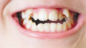
- Pericoronitis: The Most Common Soft-Tissue Red Flag
How It Presents Clinically1
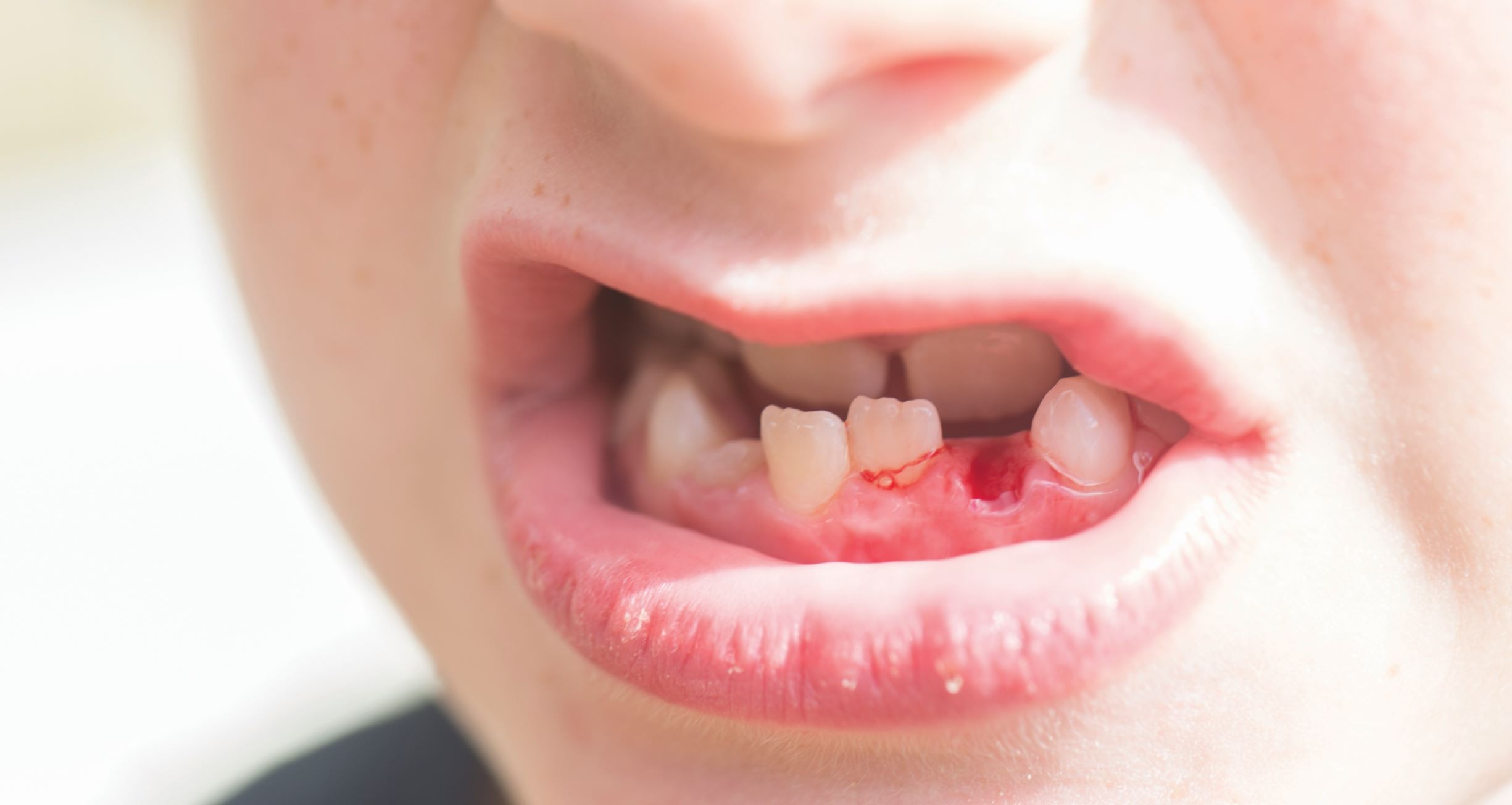
- Pain and swelling around a partially erupted mandibular third molar
- Food impaction under the operculum triggering inflammation
- Localized erythema and tenderness on probing
- Difficulty opening the mouth (mild trismus) due to inflamed opercular tissue
- Bad taste or halitosis from anaerobic bacterial buildup
- Regional lymph node tenderness in moderate cases
Epidemiological Insight 1
- A military cohort study reported a 4.92% prevalence among patients aged 20–25 years.
- 95% of infections were linked to mandibular third molars.
- Most cases appear in the 20–29 year eruption window.
Clinical Note: Recurrent pericoronitis strengthens the case for timely extraction. Partially erupted mandibular third molars with an operculum often trap food and bacteria, resulting in inflammation, pain, trismus and swelling. Operating in the presence of active infection increases surgical time, soft-tissue trauma, and postoperative discomfort.
- Typical symptoms include halitosis, purulence, and difficulty chewing.
- Stabilizing inflammation before extraction improves outcomes.
Recurrent episodes particularly indicate the need for removal.
- Age-Related Increase in Surgical Morbidity
Age isn’t just a number, it predicts how the patient will heal after surgery, which means spotting this early reduces preventable postoperative complications. Recent age‑stratified evidence shows that once patients cross 32 years 2, complication rates rise measurably.
Key Points:
- Older patient cohorts experience higher postoperative complication rates
- Delayed healing and persistent discomfort are more common after 32
- Periodontal defects distal to the second molar occur more frequently
- Increased likelihood of infection, swelling, and extended recovery time
Clinical Cue: If your patient is in—or beyond—their early 30s, delaying removal can convert a routine procedure into a morbidity‑heavy event.
As age advances, bone becomes denser and less forgiving, roots fully curve, and periodontal stability decreases. Healing slows the inflammatory burden persists longer.
- Functional Limitation: Trismus and Restricted Access
Restricted mouth opening significantly complicates surgical access, visibility, and instrumentation. Postoperative trismus is closely linked to increased inflammatory response and surgical trauma, resulting in pain, edema, and reduced inter‑incisal distance 3.
Key Points:
- Third molar surgery commonly triggers measurable reductions in mouth opening
- Inflammation and tissue trauma from flap reflection and bone removal contribute to muscle spasm
- Limited access increases operative duration and soft‑tissue manipulation, heightening postoperative swelling and discomfort
- Monitoring inter‑incisal distance helps anticipate recovery difficulty
Clinical Cue: Patients presenting with baseline restricted opening are more likely to experience prolonged postoperative discomfort and functional limitation.
- Impact on Adjacent Tooth Periodontal Health
Impacted mandibular third molars are strongly associated with periodontal defects on the distal surface of the adjacent second molar—especially when plaque retention and local inflammation persist 4.
Key Points:
- Distal bone loss and pocketing adjacent to the second molar are frequently observed
- Early intervention can prevent worsening periodontal defects
- Postoperative outcomes improve when impaction‑related inflammation is addressed promptly
- Younger patients show better periodontal recovery potential
Clinical Cue: Document baseline probing depths and bone levels. Delayed removal may lock in irreversible periodontal damage.
- Oral Hygiene and Microbial Burden
Poor plaque control around a partially erupted third molar significantly increases the risk of postoperative inflammatory complications. Elevated microbial load in the opercular region correlates with delayed wound healing, heightened inflammation, and increased pain severity.
Key Points:
- Higher pre‑operative plaque scores are associated with increased postoperative inflammation and discomfort
- Local microbial density influences surgical wound healing quality
- Mechanical debridement and pre‑operative hygiene reinforcement reduce complication rates
- Adjunctive chlorhexidine protocols may lower bacterial burden
Clinical Cue: Perform pre‑operative hygiene optimization and address local bacterial load to improve healing trajectories.
- Systemic Health Considerations
Diabetes‑related microvascular dysfunction, impaired immune response, and altered collagen metabolism can significantly delay socket healing and increase the likelihood of postoperative infection 5.
Key Points:
- Hyperglycemia impairs fibroblast activity and angiogenesis, slowing tissue regeneration
- Reduced neutrophil function increases susceptibility to oral infection
- Chronic inflammation and oxidative stress disrupt normal wound‑repair pathways
- Poor glycemic control worsens socket healing outcomes following extraction
Clinical Cue: Verify glycemic status before elective surgery. Optimize metabolic control or consider medical referrals when necessary.
Pro-Tip
Always document:
- pocket depths behind the second molar,
- presence of active infection,
- mouth-opening measurements,
- patient age-related risk discussion.
This safeguards clinical decision-making and provides medico-legal protection.
Final Thought
Third molar extraction is considered “routine,” but routine does not mean low risk. By identifying clinical red flags early, practitioners can optimize timing, minimize complications, improve periodontal outcomes, and elevate patient safety. Balancing local anatomy, systemic health, and soft-tissue status is the difference between a smooth postoperative course and preventable morbidity.
Reference
- Kwon G, Serra M. Pericoronitis.
- Yamada SI, Hasegawa T, Yoshimura N, Hakoyama Y, Nitta T, Hirahara N, Miyamoto H, Yoshimura H, Ueda N, Yamamura Y, Okuyama H. Prevalence of and risk factors for postoperative complications after lower third molar extraction: A multicenter prospective observational study in Japan. Medicine. 2022 Aug 12;101(32):e29989.
- Sreesha S, Ummar M, Sooraj S, Aslam S, Roshni A, Jabir K. Postoperative pain, edema and trismus following third molar surgery–A comparitive study between submucosal and intravenous dexamethasone. Journal of Family Medicine and Primary Care. 2020 May 1;9(5):2454-9.
- Ménager L, Ruperto M, Fricain JC, Catros S, Fénelon M. Does surgical removal of mandibular third molar influence the periodontal status of the adjacent second molars? A systematic review. Journal of Oral Medicine and Oral Surgery. 2023;29(1):1.
- Yang S, Li Y, Liu C, Wu Y, Wan Z, Shen D. Pathogenesis and treatment of wound healing in patients with diabetes after tooth extraction. Frontiers in Endocrinology. 2022 Sep 23;13:949535.
Related Contents
Video
Management of Traumatic Tooth Avulsion: When & How?
Traumatic tooth avulsion is a true dental emergency requiring immediate and structured management to...
Article
Prosthetic Design Factors That Prevent Peri-Implant Bone Loss
You placed the implant perfectly. Osseointegration was flawless. But three years later, there is 3mm...
Article
Treatment Algorithms for Peri-Implant Mucositis and Peri-Implantitis
You can prevent most peri-implant disease. But once it develops, treatment becomes less predictable....
Article
Implant Surface Decontamination: What Actually Works
Treating peri-implantitis starts with one critical step, which is decontaminating the implant surfac...
Article
Growth & Facial Asymmetry: When to Worry
Only a few dentists can notice it and realize the asymmetry can signal an underlying skeletal imbala...

Article
Orthodontic Red Flags Every Dentist Should Recognize: Functional Habits and Airway Cues
Some malocclusions cases stall for reasons you can’t see on a scan, as not...
Article
Tooth Eruption & Space Management: What Orthodontists Should Watch For
Tooth eruption may not alwaxfys fit the predictable biological timeline. A delayed or ecto...
Article
Communication & Referral Timing: Getting It Right
You have identified a problem, maybe a severe Class III malocclusion in a 9-year-old, or it is an im...